
Dr Colum Nolan
Senior Consultant Spine & Neurosurgeon
MB, BCh, BAO, LRCPSI, MRCSI, FRCSI (Neurosurgery)
Intraoperative neuroimaging in neurosurgery uses real-time imaging during surgery to guide surgeons in treating brain and spine conditions with greater precision. It helps improve accuracy, minimise risks, and ensure safer removal of tumours or correction of spinal problems.
Modern neurosurgery places strong emphasis on accuracy and adaptability, especially when operating close to the spinal cord and nerve structures. Intraoperative neuroimaging is one of the technologies used to support this approach. Unlike scans performed before surgery, intraoperative imaging allows updated images to be obtained during the operation itself, providing surgeons with real-time anatomical information.
Spine navigation and intraoperative neuroimaging are complementary technologies used in modern neurosurgery, each serving a different role during surgery. Spine navigation helps guide instruments based on mapped anatomy, while intraoperative neuroimaging provides updated images during the procedure to confirm progress and support decision-making. When used appropriately, they work together to support precision and safety during selected spine surgeries.
This page explains what intraoperative neuroimaging involves, how it is used in spine surgery, and how it supports selected treatments offered at Oxford Spine and Neurosurgery Centre.
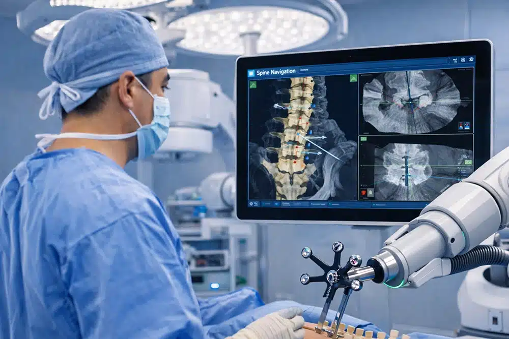
In spine surgery, intraoperative neuroimaging is often used together with navigation systems to provide updated reference points.
It may support procedures involving:
Updated imaging allows surgeons to confirm key objectives before the procedure is completed.
The effective use of intraoperative neuroimaging requires familiarity with both the technology and its integration into the surgical workflow.
At Oxford Spine and Neurosurgery Centre, intraoperative neuroimaging may be utilised where appropriate to support accuracy during neurosurgical spine procedures. Its use is guided by clinical assessment, imaging findings, and procedural requirements rather than as a routine step.
Dr Colum Nolan incorporates intraoperative imaging as part of a broader neurosurgical approach when updated visual information may add value during complex or delicate spine surgeries. This allows surgical planning to remain flexible and responsive to real-time findings.
Medical Disclaimer
This page is intended for general educational purposes only and does not constitute medical advice. Information provided is not a substitute for professional medical consultation, diagnosis, or treatment. The use of intraoperative neuroimaging depends on individual clinical assessment and suitability, as determined by a qualified medical practitioner.
Dr Colum Nolan is a Senior Consultant Neurosurgeon with over 20 years of experience, specialising in minimally invasive spine (keyhole) surgery and other spine procedures.
A graduate of the Royal College of Surgeons in Ireland, Dr Nolan underwent neurosurgical training in Ireland and Australia, followed by a fellowship in complex spine surgery at Addenbrooke’s Hospital, Cambridge, as well as rotations at the Orthopaedic Spine Unit in Norfolk and Norwich Hospital.
Dr Colum is committed to delivering compassionate, patient-centred care, combining surgical precision with a genuine dedication to improving his patients’ quality of life.

Make an Enquiry or Request an Appointment
Our friendly team is here to serve you. For urgent enquiries and appointment requests, please call or WhatsApp us directly.
